
Table of
Contents
|
Table of Contents |
This chapter covers clinical aspects of the various medical problems which may be seen in modem warfare as a result of the use of nuclear weapons. Blast, thermal, and radiation injuries are discussed first. Combined injury is discussed as a separate subject because of the special problems which patients present when radiation sickness complicates other serious injuries. The psychological and public health aspects of nuclear warfare are also combined.
The types of blast injuries by nuclear weapons are more varied than those caused by conventional weapons and are the result of two basic mechanisms, either the direct action of the blast wave overpressures or the indirect action of flying debris or violent displacement of individuals against other objects. In addition, the blast injuries caused by nuclear weapons will frequently be complicated by associated thermal and/or radiation injuries. Finally, the number of casualties produced at any one time in a given area will be very much greater for nuclear weapons than for conventional weapons.
The treatment of blast injuries is generally not difficult unless there is unrecognized internal injury with slow hemorrhage. As noted, missile injuries will predominate. About half of the patients seen will have wounds of their extremities. The thorax, abdomen, and head will be involved about equally. Missile injuries of the thorax, neck, and the head will be responsible for a large percentage of deaths because these types of injuries have a high probability of immediate fatality. The missile injuries caused by nuclear weapons will, in general, be of the low velocity type, and surprisingly severe injuries may be survived since extensive soft tissue cavitation would not be a factor. These injuries can occur with or without perforating wounds of the abdomen or the chest.
The treatment of blast injuries, whether combined with other injuries or not, is best managed by applying accepted principles of combat surgery. Treatment is divided into four basic plans:
a. Resuscitative Phase. Lifesaving resuscitative measures designed to prepare the patient for definitive surgical treatment come first. These include the establishment of the airway assuring the adequacy of respiration, replacement of lost blood and fluids, and splinting of possible fractures, particularly those involving the cervical vertebrae. Some resuscitative measures must be started prior to evacuation from the battlefield, particularly if ground transportation is used rather than helicopter evacuation.
b. Surgical Phase. Definitive surgery should be done after resuscitative measures have been used to improve the patient's condition in order to minimize the risk of surgery and anesthesia. Occasionally, lifesaving surgery must be done without delay, but normally there is time to prepare patients for surgery if they have survived long enough to reach a treatment facility.
c. Recovery Phase. In the immediate postoperative period, patients require minimal movement. Transportation to other facilities should be delayed until the patient's condition has stabilized.
d. Convalescent Phase. Patients in this phase of treatment should be evacuated back to specialized convalescent facilities in order to keep the patient load of forward surgical hospitals as low as possible. Many injuries may require a prolonged recovery period before the individual has recovered to the point where he/ she can resume their duties. Both the convalescent and recovery phases will be more protracted with the addition of the radiation injury.
Many burn casualties may occur as a result of incendiary attacks on cities and military personnel in the field during conventional warfare. However, in nuclear warfare, burns could become the most frequent injury seen. Because of the complexity of burns treatment and the increased logistical requirements associated with the management of burns, they will constitute the most difficult problem faced by the medical service.
Certain factors are of prime importance in the early evaluation of burns because of their relation to overall prognosis.
a. Area of the burns; expressed in percentage of body surface involved.
b. Involvement of critical organs; i.e., head and neck, respiratory tract, genitalia, hands, and feet.
c. Depth of burn; superficial (first- or second-degree), or deep (second degree) and full thickness (third degree).
a. The most accurate way to estimate the amount of tissue injury following a burn is to measure the extent of the body surface burned. However, direct measurement is not generally possible or necessary, and a short cut method of estimating the percent of the body surface involved can be very useful. The "Rule of Nines" method is a simple and reasonably reliable guide in which the various parts of the body are divided into surface areas of 9% each (or multiples of 9%) as shown in Table 6-I.
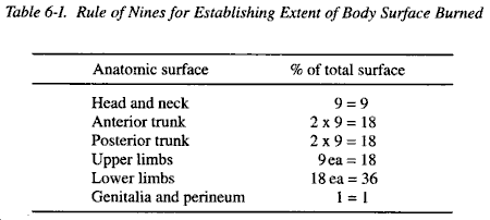
b. As the percent of the surface burned increases, morbidity and the probability of mortality increases sharply. Burns which cover 20% or more of the body surface can be fatal without treatment. Even with treatment, mortality from extensive burns will be high, particularly in the very young or the aged. Young healthy soldiers who have uncomplicated burns may survive even extensive involvement with proper care.
c. Determination of the percent of the body involved will aid in planning resuscitative treatment and estimating fluid requirements during the first 48 hours after the burn injury. Patients with severe burns will suffer quite extensive fluid and electrolyte losses, resulting in severe hypovolemic shock requiring aggressive fluid replacement therapy as early as possible. An outline of a resuscitative program is given in the treatment section.
When certain organ systems are involved, the clinical effects of burns can be quite serious in spite of the fact that only a small fraction of the body is involved.
a. Head and Neck Burns. Burns of the face can be serious problems, even if the eyes are not involved. Burns of the head frequently are complicated by severe edema, which can result in respiratory obstruction. This can be quite serious when the inhalation of hot gases has occurred. It may be necessary to do tracheotomies on many of these patients.
b. Burns of the Respiratory Tract. When hot gasses are inhaled, this very serious type of injury may be sustained. These injuries have a high probability of mortality if the burns extend deep into the alveoli. These patients are very fragile and may not tolerate early evacuation. Pulmonary edema may develop abruptly, without warning, requiring vigorous ventilator support. These injuries can be very difficult to manage.
c. Burns of Hands and Feet. These can be very disabling and may require long hospitalization for extensive surgical care even though they are not life threatening injuries. These patients may not be able to care for themselves and, as a result, will require extensive nursing care.
Burns are classified on the basis of the depth of the injury.
a. Superficial or Partial Skin Thickness Burns. These are lesions in which the dermis is intact and only the epidermis is injured. When the injury is limited and only erythema occurs (such as in a sunburn), these are usually called first-degree burns. If blistering is seen, the injuries are called second-degree burns. Superficial burns are usually painful but will heal readily by epithelization unless infection occurs. Infection can convert a typical second-degree, superficial burn into a deep or full-thickness burn which will not heal by epithelization but rather by scarring. Second-degree burns will be very common in nuclear combat and may be the one most common injury seen.
b. Deep or Full-Thickness Burns. Injuries involving the full thickness of the skin which cannot heal by epithelization are called third-degree burns. Instead, these injuries heal by scarring, and as a result there may be contraction and loss of function, particularly if extremities are involved. Extensive plastic surgery may be required to prevent or limit loss of function. The areas of a burn which are third-degree are usually painless, and this helps differentiate areas of third from second-degree when both are present. The earlier the diagnosis of the degree of burn is made, the sooner reconstructive treatment with skin grafting can be started. In general, however, in nuclear combat, early skin grafting will rarely be possible.
Initial treatment of burn patients will be resuscitative. When such patients are first seen, a simple plan of treatment must include: maintenance of airway with ventilating support as needed, adequate fluid therapy, and careful records of input and output.
a. Maintenance of Airway. This is of particular importance in head and neck burns or in unconscious patients. If large numbers of patients are seen requiring transportation over long distances early in the postburn period, tracheotomies may have to be done on a routine basis. Tracheotomies done prior to the onset of edema are much easier to perform than when they are done after edema has resulted in respiratory obstruction. When only small numbers of patients require treatment, tracheotomies are rarely required.
b. Fluid Therapy. The shock that is associated with an extensive burn will be severe, and survival of these patients depends upon adequate, balanced fluid replacement therapy. In combat, however, standardized methods of management are required. Standard formulae for determining the fluid requirements of burn patients have been developed and can be used in combat. The basic principle in these formulae is that the amount of fluid required is proportional to the percent of body surface burned and body weight. The type of fluid used includes colloidal materials to replace the plasma constituents lost as well as electrolytes.
c. Fluid Requirements for First 24 Hours.
(1) Colloid solutions: 0.5 ml x body weight in kilos x percent of body surface burned.
(2) Electrolyte solutions: 1.5 x body weight in kilos x percent of body surface burned.
(3) Additional fluids: 2000 ml 5-10% dextran in water.
d. Example. This formula, to meet the requirements of a 70-kg person with 30% body surface burn, would be:
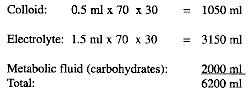
e. Restrictions. Certain restrictions on the application of this formula are required since it is only a guide.
(1) Fluid requirements for an injury involving more than 50% of the body surface should be calculated as if the burn were no more than 50%.
(2) 10,000 ml of fluid should be the maximum given in the first 24 hours.
(3) The first half of the fluid should be given more rapidly than the second; and the actual rate of administration should be 0adjusted according to urinary output.
(4) During the second 24 hours, the colloid and electrolyte given should be about one-half of that given during the first 24 hours. Again, the actual rate should be adjusted to maintain a reasonable urinary output. This is the single best clinical guide to use in determining the patient's actual fluid requirements.
(5) After the 3rd or 4th day, the patients will begin to resorb fluid from the edematous areas and will excrete it in large quantities. Administration of fluids to replace this loss is contraindicated. Excessive administration of fluids must be avoided during this time, and fluid intake can generally be reduced to that normally required for metabolic needs.
f. Input and Output Records. It is extremely important to accurately follow the input and output of fluids in burn patients. It would be impossible to modify fluid therapy according to individual needs without accurate records. Combat medical records, however, must be simple and should be attached to the patient so that they accompany him during evacuation. Medical planners must consider how to modify and improve combat medical records so that accurate input and output data on burn patients can be recorded. Most burn patients will require urinary catheterization, and this can aid considerably in recording urinary output rates accurately.
Although first priority in patient care is resuscitation, proper care of the burn wound is essential both for survival as well as for optimum healing and preservation of function. In that regard, as soon as the patient's overall condition permits, after hospitalization, initial debridement and cleaning of the burn should be done. The main purpose of this treatment is to remove foreign material and dead tissue to minimize infection. Thorough irrigation and the application of topical antimicrobial creams such as argentic sulfadiazine and sterile dressings should complete the initial procedures. Special attention should be given to critical areas such as the hands and surfaces over joints.
Radiation injury alone or in conjunction with other injuries or diseases will be common in nuclear warfare. Radiation injury can result from a single exposure to prompt radiation at the time of detonation of a nuclear weapon, from exposure to high levels of fallout radiation, or from repeated exposures to both with complex patterns of recovery from an accumulation of radiation damage.
a. Whole-body irradiation, where absorbed doses are high and acquired over short periods of time, will result in acute radiation sickness. There are three characteristic syndromes which make up the typical clinical pattern of acute radiation sickness. These are the hematopoietic, gastrointestinal, and neurovascular syndromes which occur with increasing dose respectively.
b. The hematopoietic syndrome, or syndrome of bone-marrow depression, occurs at lower doses than the others and would be the most common form of radiation sickness seen in nuclear combat. Manifestations of bone-marrow depression are seen following doses of radiation in the low through midlethal range. As the probability of lethality becomes 100 percent with higher doses, the gastrointestinal syndrome will predominate. This syndrome, which will also be common, develops from combined severe damage to bone marrow and the gastrointestinal tract. The neurovascular syndrome is associated with absorbed doses in the supralethal range and would be seen quite rarely since heat and blast effects would cause immediate lethality in most situations where the required very high radiation doses would be sustained. Exceptions could occur in aircrews exposed to prompt nuclear radiation from high altitude detonations and personnel protected against heat and blast in hardened sites below the surface or personnel in vehicles or shelters in the proximity of enhanced weapons' detonations. In these circumstances, an increase in the numbers of casualties receiving radiation doses in the supralethal range can be expected.
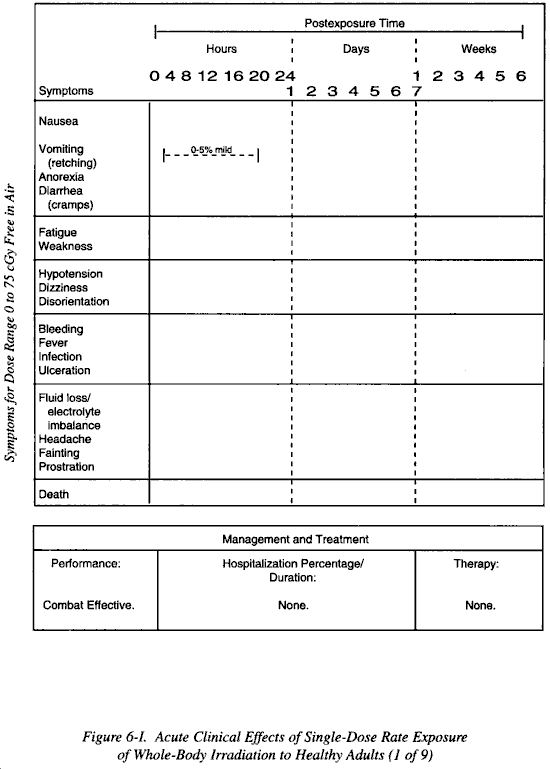
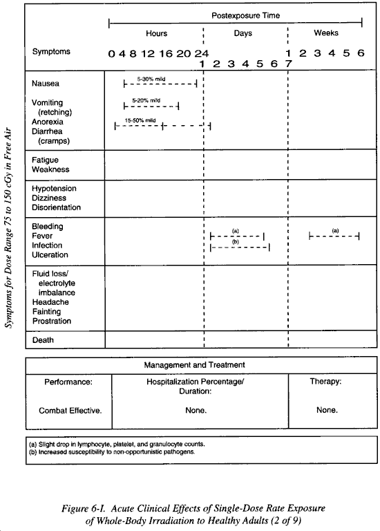
The three syndromes described follow a similar clinical pattern that can be divided into three phases: an initial or prodromal phase occurring during the first few hours after exposure; a latent phase, which becomes shorter with increasing dose; and the manifest phase of clinical illness. The time of onset and degree of the transient incapacitation of the initial phase, the duration of the latent period, as well as the time of onset and severity of the clinical phase and ultimate outcome are all to a variable extent, dose dependent.
a. Prodromal Phase. The initial phase of prodromal symptoms is characterized by the relatively rapid onset of nausea, vomiting, and malaise. This is a nonspecific clinical response to acute radiation exposure. It is not diagnostic of the degree of radiation injury; however, in the absence of associated trauma and an early onset, it does suggest a large radiation exposure. This radiogenic vomiting should not be confused with psychogenic vomiting which results from stimulation of the central nervous system by the sight/odor of blood, mutilation, vomitus, or excrement. The duration of this prodromal phase is short, generally a few hours, and the incapacitation should not be severe enough to warrant evacuation of military personnel away from their units.
b. Latent Phase. Following recovery from the prodromal phase, there will be a latent phase during which the exposed individual will be relatively symptom-free. The length of this phase varies with the dose and the nature of the later clinical phase. The latent phase is longest preceding the bone-marrow depression of the hematopoietic syndrome and may vary between 2 and 6 weeks. It is somewhat shorter prior to the gastrointestinal syndrome, lasting from a few days to a week. It is shortest of all preceding the neurovascular syndrome, lasting only a matter of hours. These times are exceedingly variable and may be modified by the presence of other disease or injury. Because of the extreme variability, it is not practical to hospitalize all personnel suspected of having radiation injury early in the latent phase unless radiation injury has reliably been diagnosed. Instead, it is much more reasonable to wait until the onset of the phase of clinical illness or the development of significant hematopoietic suppression as indicated by the individual's peripheral blood profile.
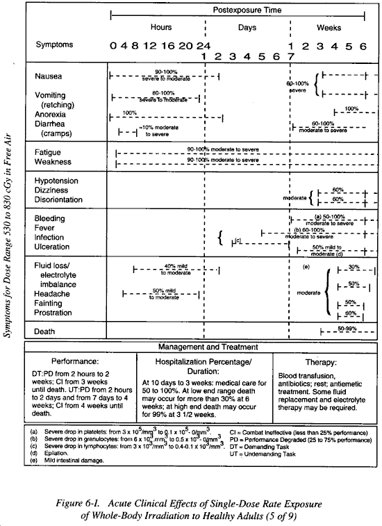
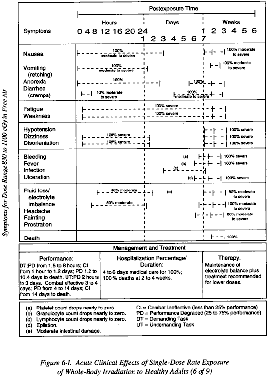
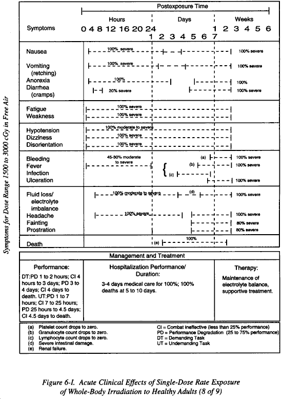
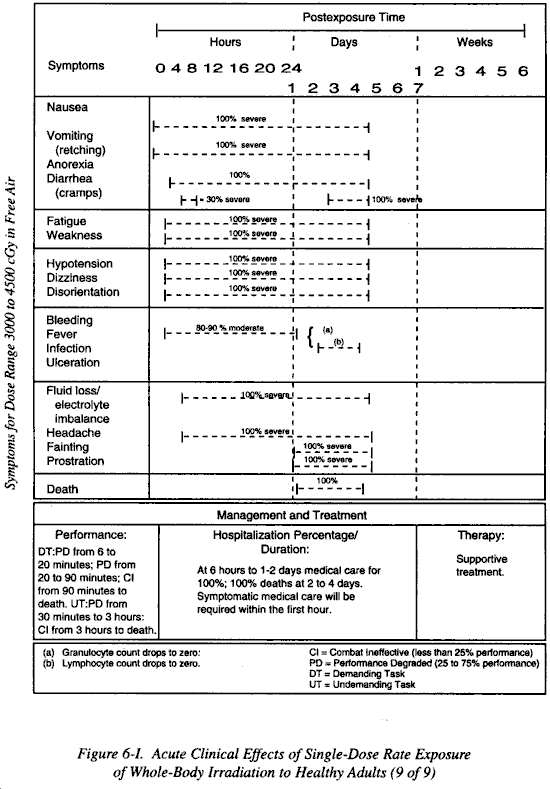
c. Manifest Phase. This phase presents with the clinical symptoms associated with the major organ system injured (marrow, intestine, neurovascular system). A summary of essential features of each syndrome and the doses at which they would be seen in young healthy adults exposed to short, high dose single exposures is shown in Figure 6-I. The details of the clinical courses of each of the three syndromes are subsequently described.
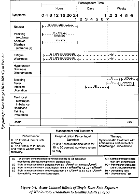
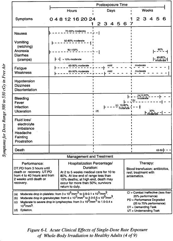
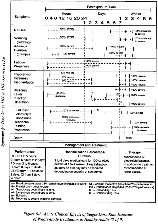
a. Patients who have received doses of radiation in the low to midlethal range will have depression of bone-marrow function with cessation of blood-cell production leading to pancytopenia. Changes within the peripheral blood profile will occur as early as 24 hours post irradiation. The exact time sequence of the depression of various circulating cell lines will vary. Lymphocytes will be depressed most rapidly and erythrocytes least rapidly. Other leukocytes and thrombocytes will be depressed somewhat less rapidly than lymphocytes. The time of onset of the depression of cellular production in the marrow will vary considerably, and the concomitant clinical problems of a tendency toward uncontrolled hemorrhage, decreased resistance to infection, and anemia will likewise vary considerably from as early as 10 days to as much as 6 to 8 weeks after exposure.
b. A reasonable average time of onset of clinical problems of bleeding and anemia and decreased resistance to infection is 2 to 3 weeks. In general, the severity of the hematological depression will be dose dependent, and the more severe phases of bone-marrow depression will occur earlier. However, even lethal cases of bone-marrow depression may not occur until 6 weeks after exposure. The presence of other injuries will increase the severity and accelerate the time of maximum bone-marrow depression
c. If the exposures leading to the bone-marrow depression are multiple, the time of onset of depression will be very difficult to estimate. The clinical picture, however, once bone-marrow depression is present, will be identical regardless of the sequence of exposure.
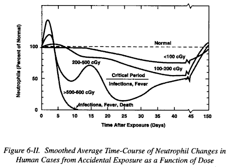
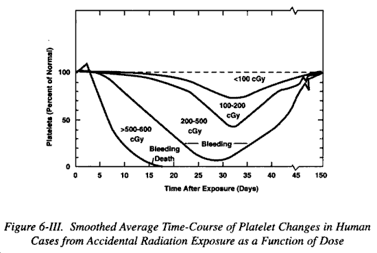
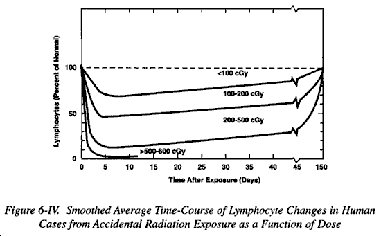
d. The most useful laboratory procedure to evaluate bone-marrow depression is the peripheral blood count. A pancytopenia with particularly severe depression of lymphocytes, granulocytes, and thrombocyte will be strongly indicative of radiationinduced bone-marrow depression. (See Figures 6-II, 6-III, and 6-IV.) Bone-marrow studies will rarely be possible under field conditions and will add little information to that which can be obtained from a careful peripheral blood count.
e. Patients will show signs or increased evidence of hemorrhagic disease and increased susceptibility to infection. If an infection occurs, there may be little clinical response because of the concomitantly depressed inflammatory response. The patients will lose weight, may lose hair and ultimately die from overwhelming infection and hemorrhage unless sufficient regeneration of the marrow occurs. Following lethal exposures, the marrow may be so damaged that recovery will be impossible.
a. The doses of radiation which will result in the gastrointestinal syndrome are higher than those causing the hematopoietic syndrome. An acute dose which will cause this syndrome would be at least 800 cGy measured in air. Under certain circumstances, lower doses may cause this syndrome, and conversely, exposures to high doses at low dose rates or as fractionated exposures may not cause it. Regardless of the dose involved, the gastrointestinal syndrome has a very serious prognosis because it is almost always accompanied by nonrecoverable bone-marrow.
b. The onset of the clinical phase of the gastrointestinal syndrome occurs earlier than that of the hematopoietic syndrome. After a short latent period of a few days to a week or so, the characteristic severe fluid losses, hemorrhage and diarrhea begin. The pathologic basis for this syndrome is an early physiologic derangement of the epithelial cells followed by a combination of severe loss of intestinal mucosa and injury to the fine vasculature of the submucosa. There is no specific clinical sign which is pathognomonic of radiation-caused gastrointestinal damage. However, a peripheral blood count done on these patients should show an early onset of a severe pancytopenia occurring as a result of the bone-marrow depression.
c. A problem in diagnosis will arise in patients with sublethal hematopoietic depression due to radiation and diarrhea due to some other cause such as infection. It would be difficult to differentiate patients with lethal radiation sickness from those with potentially nonlethal radiation sickness complicated by dysentery. Microscopic examination of the diarrhea may reveal inflammatory cells which is suggestive of dysentery. Radiation enteropathy is not likely to result in an inflammatory response. It must be assumed during the care of all patients that even those with a typical gastrointestinal syndrome may be salvageable, until blood counts indicate that the bone-marrow depression is irreversible.
This syndrome is associated only with very high acute doses of radiation. The lower limit is probably 2000 to 3000 cGy, although hypotension (significant decline in systemic blood pressure) may be seen at even lower doses. The latent period is very short varying from several hours to 1 to 3 days. The subsequent clinical picture is basically that of a steadily deteriorating state of consciousness with eventual coma and death. Convulsions may or may not occur. There may be little or no indication of increased intracranial pressure. Because of the very high doses of radiation required to cause this syndrome, personnel close enough to a nuclear explosion to receive such high doses would generally be well within the range of 100% lethality due to blast and thermal effects. However, in nuclear detonations above the atmosphere with essentially no blast, very high fluxes of ionizing radiation may extend out far enough to result in high radiation doses to aircraft crews. Such personnel could conceivably manifest this syndrome, uncomplicated by blast or thermal injury. Personnel protected from blast and thermal effects in shielded areas could also sustain such doses. Still, very few patients will be hospitalized with this syndrome.
a. The diagnosis of radiation sickness is based primarily upon the clinical picture presented by the patient. A precise history of exposure may be very difficult to obtain, since many individuals may not know that they actually have been exposed to radiation, particularly if the exposure is due to fallout. The physical findings and characteristics of the various forms of radiation sickness are described below, along with such laboratory findings as may occur. Dosimetry, at the present time, will not give adequate information to determine either the extent of radiation injury or the prognosis. Dosimeters cannot tell whether a radiation exposure is whole body or partial body. They do not tell what the dose rate of the exposure was. Finally, they cannot differentiate between single exposures and multiple exposures unless read at regular intervals.
b. These unknowns, coupled with the marked effects of age or physical condition, of concomitant disease, and of stress, etc., make it essential that physicians with the responsibility for treating patients in a hospital, base their treatment decisions primarily upon the actual clinical condition of the patient. However, in the mass casualty situation, decisions based on dosimetric data alone may be all that is practicable.
c. Consequently, the following guidelines based on recent recommendations apply to medical personnel operating in austere field conditions. Lymphocyte levels may be used as a biologic dosimeter to confirm the presence of pure radiation injury but not in combined injuries. If the physician has the resources of a clinical laboratory, additional information can be obtained to support the original working diagnosis by the presence of prodromal symptoms. An initial blood sample for concentrations of circulating lymphocytes should be obtained as soon as possible from any patient classified as "Radiation Injury Possible" or "Radiation Injury Probable." After the initial assessment or at least no later than 24 hours after the event in question, additional blood samples should be taken for comparison. The samples may be interpreted as follows:
(1) Lymphocyte levels in excess of 1500/mm3 (cubic millimeters). The patient most likely has not received a significant dose that would require treatment.
(2) Lymphocyte levels between 1000 and 1500/mm3. The patient may require treatment for moderate depression in granulocytes and platelets within 3 weeks postexposure.
(3) Lymphocyte levels between 500 and 1000/mm3. The patient will require treatment for severe radiation injury. The patient should be hospitalized to minimize the complications from hemorrhage and infection that will arise within 2-3 weeks postexposure.
(4) Lymphocyte levels of less than 500/mm3. The patient has received a radiation dose that may prove fatal. The patient needs to be hospitalized for the inevitable pancytopenic complications.
(5) Lymphocytes not detectable. The patient has received a superlethal radiation dose, and survival is very unlikely. Most of these patients have received severe injuries to their gastrointestinal and cardiovascular systems and will not survive for more than 2 weeks.
(6) Other Guidelines. A useful rule of thumb is, if lymphocytes have decreased by 50% and are less than 1000/mm3, then the patient has received a significant radiation exposure. In the event of combined injuries, the use of lymphocytes may be unreliable. Patients who have received severe burns or multisystem trauma often develop lymphopenia.
d. It is difficult to establish an early definitive diagnosis. Therefore, it is best to function within a simplified, tentative classification system based on the three possible categories of patients noted in Table 6-II and discussed in the following.
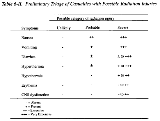
(1) Radiation Injury Unlikely. If there are no symptoms associated with radiation injury, patients are judged to be at minimal risk for radiation complications. These patients should be triaged according to the severity of the conventional injuries. If the patients are free of conventional injuries or disease states that require treatment, they should be released and returned to duty.
(2) Radiation Injury Probable. Anorexia, nausea, and vomiting are the primary prodromal symptoms associated with radiation injury. Priority for further evaluation will be assigned after all life-threatening injuries have been stabilized. Casualties in this category will not require any medical treatment within the first few days for their radiation injuries. Evidence to support the diagnosis of significant radiation injury in the absence of burns and trauma may be obtained from lymphocyte assays taken over the next 2 days. If the evidence indicates that a significant radiation injury was received, these casualties need to be monitored for pancytopenic complications.
(3) Radiation Injury Severe. These casualties are judged to have received a radiation dose that is potentially fatal. Nausea and vomiting will be almost universal for persons in this group. The prodromal phase may also include prompt explosive bloody diarrhea, significant hypotension, and signs of necrologic injury. These patients should be sorted according to the availability of resources. Patients should receive symptomatic care. Lymphocyte analysis is necessary to support this classification.
e. These symptoms frequently occur in whole-body irradiated casualties within the first few hours of postexposure.
(1) Nausea and Vomiting. Nausea and vomiting occur with increasing frequency as the radiation exceeds 100-200 cGy. Their onset may be as long as 6-12 hours postexposure, but usually subside within the first day. The occurrence of vomiting within the first 2 hours is usually associated with a severe radiation dose. Vomiting within the first hour, especially if accompanied by explosive diarrhea, is associated with doses that frequently prove fatal. Due to the transient nature of these symptoms, it is possible that the patient will have already passed through the initial phase of gastrointestinal distress before being seen by a physician. It will be necessary to inquire about these symptoms at the initial examination.
(2) Hyperthermia. Casualties who have received a potentially lethal radiation injury show a significant rise in body temperature within the first few hours postexposure. Although the number of cases is few, this appears to be a consistent finding. The occurrence of fever and chills within the first day postexposure is associated with a severe and life-threatening radiation dose. Hyperthermia may occur in patients who receive lower but still serious radiation doses (200 cGy or more). Present evidence indicates that hyperthermia is frequently overlooked. Individuals wearing a chemical ensemble will normally be hyperthermic; consequently, this will not be a useful sign.
(3) Erythema. A person who received a whole-body dose of more than 1000-2000 cGy will develop erythema within the first day postexposure. This is also true for those who received comparable doses to local body regions, when the erythema is restricted to the affected area. With doses lower but still in the potentially fatal range (200 cGy or more), erythema is less frequently seen.
(4) Hypotension. A noticeable and sometimes clinically significant decline in systemic blood pressure has been recorded in victims who received a supralethal whole-body radiation dose. A severe hypotensive episode was recorded in one person who had received several thousand rads. In persons who received several hundred rads, a drop in systemic blood pressure of more than 10% has been noted. Severe hypotension after irradiation is associated with a poor prognosis.
(5) Necrologic Dysfunction. Experience indicates that almost all persons who demonstrate obvious signs of damage to the central nervous system within the first hour postexposure have received a superlethal dose. Symptoms include mental confusion, convulsions, and coma. Intractable hypotension will probably accompany these symptoms. Despite vascular support, these patients succumb within 48 hours.
f. Casualties who have received a potentially fatal dose of radiation will most likely experience a pattern of prodromal symptoms that is associated with the radiation exposure itself. Unfortunately, these symptoms are nonspecific and may be seen with other forms of illness or injury, which may complicate the process of diagnosis. Therefore, the triage officer must determine the symptoms that have occurred within the first day postexposure, evaluate the possibility that they are indeed related to radiation exposure, and then assign the patient to one of the three categories: "Radiation Injury Unlikely"; "Radiation Injury Probable"; "Radiation Injury Severe." In the last two categories, the study of changes in circulating lymphocytes may either support or rule out the original working diagnosis. All combined-injury patients should be treated initially as if no significant radiation injury is present. Triage and care of any life-threatening injuries should be rendered without regard for the probability of radiation injury. The physician should make a preliminary diagnosis of radiation injury only for those patients for whom radiation is the sole source of the problem. This is based on the appearance of nausea, vomiting, diarrhea, hyperthermia, hypotension, and necrologic dysfunction.
a. Radiation injury per se does not imply that the patient is a health hazard to the medical staff. Studies indicate that the levels of intrinsic radiation present within the patient from activation (after exposure to neutron and high-energy photon sources) are not life-threatening.
b. Patients entering a medical treatment facility should be routinely decontaminated if monitoring for radiation is not available. Removal of the patient's usually reduce most of the contamination. Washing exposed body clothing will surfaces will further reduce this problem. Both of these procedures can be performed in the field or on the way to the treatment facility. Once the patient has entered the treatment facility, care should be based on the obvious injuries. Care for life-threatening injuries should not be delayed until the decontamination procedures are completed.
c. When radiation safety personnel are available, decontamination procedures will be established to assist in rendering care and to minimize the hazard from radioactive contaminants. A more extensive decontamination procedure is to scrub the areas of persistent contamination with a mild detergent or a diluted strong detergent. Caution should be taken to not disrupt the integrity of the skin while scrubbing because disruption can lead to incorporation of the radioisotopes into deeper layers of the skin. Contaminated wounds should be treated first, since they will rapidly incorporate the contaminant. Washing, gentle scrubbing, or even debridement may be necessary to reduce the level of contaminants.
d. Wearing surgical attire will reduce the possible contamination of health personnel. If additional precautions are warranted, rotation of the attending personnel will further reduce the possibility of significant contamination or exposure. The prevention of incorporation is of paramount importance. The inhalation or ingestion of radioactive particles is a much more difficult problem, and resources to deal with it will not be available in a field situation.
a. The primary determinants of survival among most patients receiving intermediate (serious but not uniformly fatal if treated) radiation doses is management of microbial infections and stopping any bleeding. If high intermediate doses have been received, fluid and electrolyte loss may cause early deaths. If properly resuscitated, however, these patients may survive until the consequences of hematologic failure become apparent.
b. For those casualties who have received sublethal whole-body radiation doses, gastrointestinal distress will predominate in the first 2 days. Antiemetics (metocloproparnide, dazopride) may be effective in reducing the symptoms, but present drugs available have significant side effects. Unless severe radiation injury has occurred, these symptoms will usually subside within the first day. For those patients who continue to experience gastrointestinal distress, parenteral fluids should be considered. If explosive diarrhea occurred within the first hour postexposure, fluids and electrolytes should be administered if available. For triage purposes, the presence of explosive diarrhea (especially bloody) is likely to be related to a fatal radiation dose.
c. Cardiovascular support for patients with clinically significant hypotension and necrologic dysfunction should be undertaken only when resources and staff allow. These patients are not likely to survive injury to the vascular and gastrointestinal systems combined with marrow aplasia.
620. Diagnosis and Treatment of Patient With Combined Injuries.
a. Conventional injuries should be treated first, since no immediate life-threatening hazard exists for radiation casualties who can ultimately survive. The patient with multiple injuries should be resuscitated and stabilized. During this process standard preparation for surgery will accomplish much radioactive decontamination. After surgery more definitive evaluation of radiation exposure can be initiated.
b. In the event of a radiation accident or nuclear detonation, many patients will probably suffer burns and traumatic injuries in addition to radiation. The initial triage of combined injury patients is based on these conventional injuries. Further reclassification may be warranted on the basis of prodromal symptoms associated with radiation injury. The prognosis for all combined injuries is worse than for radiation injury alone. Animal studies indicate that when other injuries are accompanied by sublethal doses of radiation, infections are much more difficult to control, and wounds and fractures heal more slowly. Thus, potentially survivable burns and trauma will be fatal in a large percentage of persons who have also received significant injury from sublethal doses of radiation. Often with conventional injuries, staged reparative surgery is scheduled for 1-2 days after the initial surgery, and reconstructive surgery is still later. Because of the delays in wound healing and the subsequent granulocytopenia and thrombocytopenia with injuries from nuclear weapons, most of the life-saving and reconstructive surgery must be performed within 36 hours after the exposure. Then, if possible, no surgery should be performed for the next 1 1/2-2 months postexposure.
a. In spite of antibiotics, infections with opportunistic pathogens are still a major problem. The majority of these organisms today are gram-negative like Escherichia coli, Pseudomonas aeruginosa, and many others. These infections occur as a consequence of both profound immunosuppression and abnormal colonization of body surfaces and invasive medical devices. Susceptible body surfaces include the oropharyngeal-respiratory tree and the intestine. Wound sites and artificial invasive devices such as catheters are also important sources of infection. Infections may be more prevalent and severe if patients are maintained for long periods in environments containing antibiotic resistant pathogens.
b. Wound debridement, dressings, and, when necessary, antibiotics are key elements in infection control. Antibiotics, preferably in appropriate combination in therapy, should be used promptly to treat any new fever. When signs or symptoms of infection do appear in the granulocytopenic patient, treatment should be started without waiting for culture and sensitivity studies. Initial coverage should include gram-negative organisms and Staphylococcus aureus. Prevalent organisms and antimicrobial susceptibility patterns in the particular medical facility should also be considered. The drugs most often used now for the initial treatment are the synthetic penicillins, such as ticarcillin, combined with an aminoglycoside like tobramycin. It is recommended either that the treatment continue until the granulocytes return to more than 500 or treat for just 2 weeks and stop even if the white cell count is still low, as long as all signs of infection have cleared.
c. Systemic antibiotic therapy for management of infection is as follows.
(1) Types of Agents.
(a) Aminoglycosides such as gentarnicin, netilimicin, tobramycin, and amikacin are the most effective.
(b) Ureidopenicillins and carboxypenicillins such as ticarcillin and peperacillin are less effective than the aminoglycosides, but are synergistic with them against gram-negative enterics.
(c) Monobactams are effective against gram-negative enterics, to a lesser degree then aminoglycosides, but have no renal toxicity as they do.
(d) Beta Iactam resistant penicillins such as methicillin or dicloxicillin are effective for therapy of Staphylococcus aureus. Vancomycin can be administered for therapy of methicillin resistant S. aureus.
(e) Irnipenem (combined with cilastalin) is the only single agent that is effective against aerobic gram-positive and gram-negative organisms as well as anaerobic bacteria. However, some strains of Pseudomonas may be resistant.
(2) Combination Therapy. Several combinations have been advocated for the therapy of mixed aerobic-anaerobic infection, or for the therapy of gram-negative infections in the compromised host.
(a) Gram-negative infection: Aminoglycoside plus ureidopenicillins or carboxypenicillins; aminoglycoside plus a cephalosporin (second or third generation; arninoglycoside plus a monolactam.
(b) Gram-positive infection: Combinations of beta lactim resistant penicillin and an aminoglycoside.
(c) Mixed aerobic-anaerobic infections: An arninoglycoside or quinoline plus either clindamycin, cefoxitin, or metronidazole.
a. Treatable radiation-associated injuries only include those with the hematologic and possibly, the gastrointestinal syndrome. Combined injuries would shift the treatable range of injuries to the lower radiation doses. Even in these ranges there is very little definitive information available now. Many approaches suitable for conventional injuries may be found of little utility in irradiated subjects.
b. First actions in dealing with radiation casualties are to treat any conventional injuries first. Maintain ventilation and perfusion and stop hemorrhages. Most decontamination will be accomplished through routine management of the patient. Triage for radiation injuries followed by steps to prevent infection, fluid and electrolyte imbalance and bleeding will be necessary after patient stabilization. Unfortunately, there are limitations in the ability to effect these treatments successfully, particularly on a large scale with limited resources.
c. Presently new means of radioprotection and repair of radiation damage are on the horizon. Furthermore, immunomodulators are now under study which may not only facilitate marrow regeneration, but also help reduce the profound immunosuppression responsible for infections associated with severe injury. These agents may be used in combination with radioprotectors and antibiotics to further enhance survival. Leukopenia is a significant problem in irradiated casualties, but hazards exist with the transfusion of leukocytes into patients. Stem cell regeneration into selected populations probably offers the best opportunity to correct this deficiency. Although platelet transfusions are certainly desirable for radiation victims, they are presently not practical for mass casualty scenarios. A similar situation exists for bone marrow transplantation, although enormous progress is being made in autologous transplants. Again, stimulation of repair of surviving stem cells is probably the best near term hope of solving this problem. Problems of effective wound management and fluid and electrolyte replacement remain to be overcome in the neutropenic patient. Pharmacologic means to regulate performance decrements such as emesis and early transient incapacitation still are not available for use by military personnel.
d. The foregoing should clearly show that much remains to be done to achieve effective treatment of radiation or combined injury victims. However, progress in this area is being made and the concerns outlined above will be resolved.
a. At Hiroshima and Nagasaki, large numbers of patients with traumatic injury developed complications 2 to 3 weeks after exposure which were characteristic of the effects of bone-marrow depression. The open wounds of many patients stopped healing and became hemorrhagic. There was an associated loss of granulation tissue. Patients lost weight, and many died as a result of overwhelming sepsis. Those patients who recovered went onto normal wound healing after return of bone-marrow function. This would be the typical clinical picture in patients exposed to prompt radiation from small weapons, while at the same time sustaining thermal or blast injuries. The most common form of radiation sickness would be the hematopoietic syndrome, and the resultant hemorrhagic tendencies and decreased resistance to infection would complicate the healing of these patients' wounds. The overall result would be prolonged hospitalization and increased morbidity and mortality.
b. Unfortunately, it will not always be possible at the time of admission to predict which patients with thermal or blast injuries would develop radiation sickness. A history of the prodromal symptoms which typically follow radiation exposure, as described previously, would be helpful but could not be relied upon. The first reliable indication that complications of radiation sickness might occur would be a lymphocytopenia, neutropenia, and thrombocytopenia noted in the peripheral blood count. By that time, however, the patient should have had at least the initial surgery required for his or her primary injuries. Subsequently, during the time the patients would be in the clinical phase of bone-marrow depression, careful supportive therapy would be required and elective surgical procedures should be avoided. Only those procedures that are actually required to save life and limb would be indicated. If surgery is required during the clinical phase of radiation sickness, increased morbidity and mortality would be expected. This could be minimized by applying the basic techniques of meticulous surgical care such as are commonly used in noncombat surgery on patients with hemorrhagic disorders.
c. Patients will also be seen who will have sustained their traumatic injuries and their exposures to radiation at different times. The best example of this would be patients wounded by conventional weapons before or after being exposed to fallout radiation. The interaction of the bone-marrow depression with traumatic injury is highly dependent upon this factor of timing. When patients are in the middle of the clinical phase of bone-marrow depression and are injured, the effect of this combination will be very deleterious, and a high mortality rate will be seen. If, on the other hand, the clinical phase of sickness comes late in the course of wound healing, a relatively small interaction will be seen.
d. The degree of interaction between radiation sickness and traumatic injury will also depend a great deal upon the time course of the traumatic injury. Patients with small wounds that can be closed primarily, or with closed fractures which can be immobilized early, will not be as sensitive to the effects of radiation over as long a period of time as those patients with severe open wounds or burns. burn patients, in particular, will be susceptible to infection for an extended period of time and will be particularly sensitive to the decreased resistance to infection characteristic of radiation sickness. It will be expected then that the morbidity and mortality of burns combined with radiation sickness would be much greater than the morbidity and mortality following minor closed wounds and fractures. Open wounds and extensive soft tissue injuries would behave similarly.
e. Radiation injury can be combined with a number of other clinical problems. Radiation sickness may be superimposed on underlying medical diseases, and such patients will also be more sensitive to the deleterious effect of radiation sickness. There have been indications that radiation sickness will allow otherwise nonpathogenic bacteria to become pathogenic and to cause significant disease. Further, patients with mild radiation sickness which might otherwise go unrecognized would be much more sensitive to environmental stresses or to the effects of chemical and biological agents.
a. Many factors are responsible for relative radiation sensitivity. In any given population, some individuals will naturally be sensitive to irradiation and others will be relatively resistant. The factors which determine this are genetic as well as nongenetic. Age and physical condition are very important. The general condition of the individual at the time of exposure can modify the response to radiation considerably. There may be increased resistance to radiation if an individual has been exposed to a stressful stimulus such as a minor traumatic injury or environmental stresses prior to a radiation exposure. This phenomenon has been demonstrated in a number of laboratory studies with a number of animal species and a wide variety of stresses. Whether this applies to humans and to what degree is not known, but what can be said is that in combat situations that dose of radiation which would result in a given clinical response with a given probability is almost impossible to estimate.
b. An example of this problem is the question of the LD50 for people. A specific LD50 for individuals in combat cannot be given except as a broad range. The LD50 for a young adult unstressed and subject to a single acute exposure of gamma radiation would probably be in the range of 450-500 cGy. There are indications that neutrons are more effective in producing lethality. (See paragraph 504b.) If the individual is stressed prior to radiation with a minor injury, the dose required to give a 50% probability of lethality may be increased by 50% or more. If on the other hand the radiation exposure is followed by some other injury, the dose which would result in a 50% mortality might very well be decreased by a factor of two. If an individual is exposed to a number of low dose rate, small exposures such as would occur from repeated entry into fallout fields, the dose required to result in 50% mortality would be increased.
c. If the factors of age, different physical conditions, etc., are added, and a large group of individuals are exposed to a variety of radiation exposures, combined or not combined with a variety of stresses and injuries, the result is a range for the LD50 that could be from as low as 200 to 450 cGy. This is an estimate, and proof of this will only come from actual combat experiences. If the exposure is a low dose rate exposure received over a long period of time (as in the case of fallout), the LD50 dose range could be considerably higher than 450 cGy. But variations such as this are quite possible and indicate why personnel dosimetry cannot be used as an absolute indication of prognosis. This is summarized in Figure 6-V.
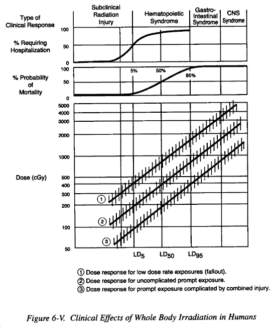
d. Dosimetry for an individual patient should only be considered as an aid to diagnosis and prognosis. The patient's clinical condition combined with appropriate laboratory investigation will indicate the prognosis much better. It is perfectly possible for patients with a total exposure of 1000 rads or more, as recorded by personal dosimeters, to survive if that exposure was accumulated over a long period of time and particularly if it is not whole body and the patient is a young healthy adult.
a. For centuries the conduct and outcome of military operations have been profoundly affected by a small number of infectious diseases. The disruptive effects of war result in conditions conducive to increases in the incidence of these diseases, often in epidemic proportions. The use of nuclear weapons, with their potential for massive destruction, would produce situations in which epidemic outbreaks of disease among civilian populations would become highly probable. Enteric and respiratory diseases would be particular problems. These, in turn, could present serious hazards to military forces in the area and serious problems for a military medical service, particularly when civilian medical facilities and personnel are inadequate to handle the problems.
b. If large, heavily populated areas are devastated, the social organization which is required to effectively support a modem medical care system will be severely compromised. It is not until society reorganizes itself and rebuilds that a complex system such as modem medical care will resume. In past wars, military medical forces have provided for civilian populations and also the means for the rebuilding of a civilization ravaged by war. If the ravages of war are beyond the capabilities of either the society itself or the armies operating in the areas to repair, then the balance will be tipped in favor of decimation of the population by the classical diseases of disaster such as dysentery, typhus, typhoid fever, cholera, and plague. The results could be devastating to modem civilization.
The concept of what has been termed "Nuclear Winter" is a rather recent concern. This is a phenomenon that has attracted much attention but little serious research. In the early 1980's the issue was politicized for various reasons. Therefore, a considerable amount of conjecture and hyperbole has surrounded the discussion of nuclear winter. However, there are certain phenomena that will be experienced in large amounts of dust, smoke, and debris injected into the upper atmosphere. This cloud would have a tendency to absorb or scatter the sunlight thus decreasing the surface temperature over a portion of the earth. This could conceivably interfere significantly with the production of foodstuffs in these regions. There is an additional concern that in the event of a high air burst, the nitrogen in the upper atmosphere would be converted into oxides and the oxides, in turn, would combine with the ozone layer thus depleting the protective ozone. This would cause a significant increase in the amount of ultraviolet light capable of reaching the earth's surface. The ozone layer would eventually be reestablished. The combination of cooling, decreased ambient light, and increased ultraviolet light bombardment could have a significant impact on food production and perhaps energy consumption. Serious research is needed to attempt to quantify these effects.
Although it is possible to estimate roughly the number of injured and dead which would result from the thermal, blast, and radiation effects of a nuclear explosion, it is much more difficult to predict the numbers and types of psychiatric patients. It is generally felt that the types of acute psychological problems which would occur in such circumstances would be essentially the same as those seen in other combat situations, and that the treatment methods which have been developed as a result of experience in past wars would be appropriate.
a. The primary psychological abnormality which develops in severe stress or disaster situations is a transient, fluid state of emotional disruption. This occurs when individuals cannot cope with the danger presented to them by their environment. Its major features are fear and the results therefrom. The fear develops largely from the individual's inability to make meaningful decisions or initiate purposeful actions; and, as a result, even minor decisions become difficult to make. A vicious circle of fear-inaction-fear may ensue, and the individual involved may become ineffective. This may vary in degree all the way from very mild impairment of effectiveness to complete helplessness. Panic, defined as frantic, irrational behavior associated with real or supposed trapping, probably would be rare, since it has been found to be rare in other disaster situations. Precipitous flight with direction and purpose is not panic and should be considered a psychologically useful and practical response to the situation.
b. The characteristic disturbances which would occur include: stunned mute behavior, uncontrolled flight, tearful helplessness, apathetic depression, inappropriate activity, increased tension, or preoccupation with somatic representations. These disturbances can last for minutes, hours, days, or sometimes weeks. Longer term reactions may include phobias, survivor guilt, and psychosomatic symptoms. Fortunately, patients with the milder and shorter disturbances are in the majority.
The frequency and severity of the psychological disturbances vary with several factors.
a. Intensity and Severity of Stress. Stressful situations of brief duration are rather easily tolerated, and recovery of individuals with mild degrees of mental disruption under these circumstances is rapid. If stressful situations follow one another rapidly, or if any one of them is of long duration, then the probability of the occurrence of more severe psychological reactions of longer duration increase.
b. Degree of Personal Involvement. If individuals have "close calls" or if they see close friends or relatives severely injured, their reactions will be more severe than if they remain relatively remote from danger.
c. Degree of Training. This is the most important factor in that it is one which is most easy to modify. Well-trained individuals, who can react readily to dangerous situations and initiate appropriate actions, will develop a minimum of incapacitating fear. The fear they do develop will, if anything, help them, since it will bean integral part of a reaction of increased awareness or alertness allowing more efficient fight or flight.
d. Degree of Warning. This is closely related to the above. Warning helps trained persons to prepare. They can initiate proper actions early. For untrained persons, the effect will be variable. If the fear is not incapacitating, then untrained persons who cannot react automatically to initiate proper actions may be able to utilize the time to improvise appropriate action. Whatever time they have to do this will help.
e. Presence of Leadership. In a disaster situation, a few individuals will emerge as leaders in a group. These may not be the appointed leaders, although in a military unit this is usually not the case unless the appointed or regular leaders become ineffective or are lost. When effective leadership is available, the group will fare much better than when there is none.
f. Group Identification. This is a particularly important factor for the military. If group or unit integrity is preserved, the individuals in the unit will do much better. Also, those individuals with mild psychological disruptions will recover faster if they can remain with or close to their unit, thus retaining their personal relationship as a member of the unit.
a. A major characteristic of these patients will be their suggestibility, and it is this which forms one of the basic underlying principles of treatment. The psychological disorders described do not require elaborate treatment and the best treatment is that which is simple, direct, and immediate. It should be done as far forward as possible, preferably within the unit to which the individual belongs. If this is not possible, then it should be started as soon as possible and in a medical facility close to the individual's unit. Evacuation to distant medical facilities is contraindicated. Evacuation tends to make the psychological problems worse by severing the patient's relationship with his or her group or unit and by introducing the element of "secondary gain" with the removal of the patient from danger.
b. Treatment consists of:
(1) Reassurance and suggestion that the situation will improve. These people are suggestible early in their disruptive phase and simple reassurance using a positive, direct approach is usually successful. The individual should be made to feel that he or she has an excellent chance of recovery, which, in general, is true.
(2) Rest with removal from immediate danger. A short period of rest in a safe area is of great benefit.
(3) Catharsis. Retention of fear and anxiety by the more severely incapacitated frequently blocks effective communication: When the patient expresses his or her feelings, this tends to remove the block. This communication is essential before the individual can recover enough to rejoin the activities of his or her group or unit.
c. Psychiatrists are not always available to participate in the overall treatment of such patients. Therefore, all medical officers and their staffs should be familiar with these principles for managing the psychological problems arising from such disasters. The success of their actions will depend largely on how well the line commanders understand the program of managing this problem, since in a great degree the practical therapy of the mildly affected will be, in fact, the positive leadership actions taken by commanders.
The most important preventive factor is intensive training. The end result is less fear and more prompt effective action. Action relieves tension so that the fear response is less likely to become severe or incapacitating. Fear may not even develop to the point where the individual is aware of it. Other factors which contribute to prevention include discipline, morale, good leadership, and promotion of group identification. The beneficial results of effective command cannot be overemphasized.