Table of
Contents
|
Table of Contents |
Paragraph A.01. Introduction
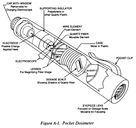
Figure A-I. Pocket Dosimeter
a. The detection and repeated measurement of radioactive fallout fields produced by nuclear explosions will give important information affecting the operation of field medical units. In addition, medical personnel must be trained in the detection and measurement of patient contamination in order to prevent uncontrolled exposures of hospitals and personnel.
b. The purpose of this Annex is to provide medical personnel with information on the basic principles of radiation dosimetry, the operation of radiation detection instrumentation, and the techniques used in performing radiological surveys.
This Annex discusses some of the operating principles and applications of various radiation detection and measurement instruments that may be used by the military. Definitions and descriptions of terms used in connection with radiation detection and measurement instruments are presented first. The operating principles and applications of selected instrumentation are then described. Finally, basic techniques for performing radiological surveys are summarized.
a. Radiation Absorbed Dose (rad). The term "rad" is an abbreviation for radiation absorbed dose and is a measure of the energy deposited in matter by ionizing radiation. The International System SD (skin dose) unit for radiation absorbed dose is the Gray (Gy), (1 Joule per kilogram), 1 Gy = 100 rad; 1 centigray (cGy) = 1 rad. The cGy is a measure of absorbed dose in any material. Use of the term cGy is not restricted to x- or gamma radiation, but can be used for all forms of ionizing radiation, including particulate. Dose means the total amount of energy absorbed. The exposure could be single or multiple and either short or long in duration.
b. Dose Rate. Dose rate is the dose of radiation per unit time.
c. Excitation. Excitation is a change in energy level of an orbital electron and occurs when the energy lost by the incident radiation is insufficient to cause ionization. It can result from interactions involving incident photons of gamma or x-radiation or from inelastic collisions of particles. When the excited electron returns to its ground-state energy level, it must give off energy in the form of a photon of a electromagnetic radiation, which is usually of low enough energy to be detected with a photomultiplier tube.
d. Ionization. Ionization is the separation of an electrically neutral atom or molecule into electrically charged components termed an ion pair. This is accomplished by removal of one or more electrons (negatively charged) from an atom or molecule which then becomes a positively charged ion.
e. Specific Ionization. Specific ionization is the number of ion pair per unit distance formed along the path of a particle. The distance specified is usually centimeters. The density of ionization produced by a given particle is a function of its charge and its velocity. A more slowly moving particle spends more time in the vicinity of the atom or molecule, increasing the chance of ionization occurring. As an example, because of its slowness and charge, an alpha particle will produce thousands more ion pairs per cm of travel than an electron (beta particle) of the same energy (approximately 100 ion pairs per cm).
f. Gas Amplification. The gas amplification factor is defined as the ratio of the number of electrons collected by the anode to the number of electrons formed by the primary radiation interaction. If sufficient potential difference is applied across a detector's electrodes, the free electrons resulting from ionization are accelerated toward the anode with enough energy to ionize the neutral gas atoms that are in their path, resulting in secondary ionization. This secondary ionization or amplification in the gas can add many thousands of free electrons to the sensitive volume for each primary electron that was formed by the radiation.
g. Pulse Height. Pulse height, which is a quantitative measurement of current flow, depends upon both the gas amplification factor and specific ionization of a given radiation. Since alpha particles have the highest specific ionization, they will produce the largest pulses. Beta particles, having a specific ionization of about 1/1000 that of alpha particles of equivalent energy, will produce pulses about 1/1000 the amplitude of those produced by alpha particles.
h. Free in Air Dose. The term "free-in-air-dose" refers to the radiation which would be measured in air at a certain point. This differs from other radiation doses of importance that might actually be desirable to measure, such as midline tissue dose or dose to the blood forming organs. The differentiation is made since free-in-airdose is exceedingly easy to measure with current field instruments, while the more meaningful dose may be estimated or determined by use of a scaling factor or conversion approximation. Military tactical dosimeters measure free-in-air-doses.
a. Human senses do not respond to ionizing radiation. Accordingly, special instrumentation must be used for radiation detection and measurement. Since the degree of hazard from radiation to humans depends on the type of radiation, its energy spectrum, as well as the quantity to which a person has been exposed, radiation detectors used in the field must be capable of making qualitative as well as quantitative measurements.
b. An ideal instrument for practical use in the field would have the following characteristics:
(1) Measure dose, or dose rate, in units directly applicable to the tissue of concern.
(2) Respond to only one kind of radiation at a time.
(3) Have the desired sensitivity and accuracy, independent of the energy of the radiation.
(4) Be free of calibration and zero drift within reasonable times.
(5) Be free of extraneous effects from gravitational forces, temperature, humidity, chemical vapors, electrical and magnetic fields, shock, and so on.
(6) Have a short response or readout time.
(7) Indicate the occurrence of full- or off-scale readings if its range is exceeded.
(8) Have a means of field checking its operability.
(9) Have a means of indicating when it is inoperable.
(10) Be small, light, and rugged, particularly if it is a portable instrument.
(11) Be relatively trouble-free and require only infrequent, simple repair and maintenance.
c. No single instrument at present has all the characteristics described. Accordingly, different types of instruments must be used depending upon the nature of the radiation hazard. The characteristics of some of the more commonly used detectors are summarized below.
(1) Ionization Chambers. Ionization chambers measure dose and dose rate from gamma and x-radiations. A typical ionization chamber that measures total dose is the pocket dosimeter. It is the size of a large fountain pen. (See Figure A-I.) It has a chamber containing two electrodes, one of which is a quartz fiber loop free to move with respect to its mounting. Radiation entering the chamber causes ionization within the sensitive volume. The distance the fiber moves is proportional to the dose received in the chamber. Instruments of this type are sensitive to severe shock and humidity and small enough to be worn comfortably. The great advantage of this type of instrument illustrated, is that it can be read at any time without the aid of a supplementary charger-reader by simply holding it up to a source of light and looking into it.
(2) Geiger-Mueller Counter. Geiger-Mueller counters are normally used for detecting single ionizing events which take place within the sensitive volume of the counter. They are very rugged and sensitive to low levels of radiation. They are usually equipped with audible detection of radiation in the form of "clicks." Geiger-Mueller counters detect gamma photons or beta particles. Detection of gamma rays is less efficient than of beta particles. A discriminating shield is usually provided with Geiger-Mueller instruments which when opened admits both beta and gamma radiation. With the shield closed, only gamma is admitted. Use of the shield may permit qualitative differentiation between ionization caused by beta particles and that produced by gamma photons.
(3) Proportional Counters. Proportional counters are used to detect one type of radiation in the presence of other types of radiation or to obtain output signals greater than those obtainable with ionization chambers of equivalent size. Proportional counters may be used to either detect events or to measure absorbed energy (dose), because the output pulse is directly proportional to the energy released in the sensitive volume of the counter. Proportional counters are most widely used for the detection of alpha particles, neutrons, and protons.
(4) Scintillation Counters. A scintillation counter combines a photomultiplier tube with a scintillating material which may be a crystal or other phosphor (solid, liquid, or gas). Light pulses produced in the scintillator by radiation, releases photoelectrons from the cathode of the photomultiplier tube, which then initiates pulses of current that can be counted. Scintillation counters are available that can detect alpha and beta particles, gamma rays, neutrons, protons, and electrons. The most common for field use are those employed as alpha counters or as gamma detectors. Although very energy dependent, scintillation counters are more efficient at detecting low level gamma backgrounds than are Geiger-Mueller counters.
(5) Chemical Dosimeters. Chemical dosimeters are systems in which measurable chemical changes are produced by ionizing radiation. Radiation produces acids in the system, the amount of which can be determined from visible color changes, or, more accurately, by titration or pH readings. Most chemical systems of practical size are useful only for gamma doses of hundreds to millions of cGy. However, small volume detectors can be found which measure doses in the range of a few cGy to several thousand cGy.
(6) Photographic Emulsions. Photographic emulsions are frequently used as detectors. The film badge has been the most common dosimeter in use, but is tending to be replaced by thermoluminescent dosimeters (TLD). The film badge uses the effect of radiation on photographic film to record dose. After film developing, the optical density is compared to a film calibration curve, and a measure of exposure dose is obtained. As the exposure dose increases, the optical density of the emulsion increases. At least two different types of film are employed to cover a wide-exposure range; a low exposure film, 0.02 cGy to 2 cGy and a high-exposure film, 1 cGy to 1,000 cGy. Metal filters such as aluminum, copper, and cadmium-tungsten, are used to increase the accuracy in the reading. The heavy metal filter also intensifies the gamma radiation interaction. Beta radiation is evaluated by observing the density change to a portion of film which is not covered by a filter. Film badges or TLDs are widely used as they provide an accurate means of recording radiation exposure at a low cost. Their disadvantage is that heat, moisture, and aging will cause a natural change in the films optical density.
(7) Thermoluminescent Dosimeters. Thermoluminescent dosimeters (TLDs) detect radiations by the formation of a metastable crystalline structure with the valence electrons. Ionizing radiation excites valence electrons to a state within the crystal structure which can be detected by heating the crystal. When heated, the electrons are released from these traps and return to their lowest energy state with the release of light. The amount of light released is proportional to the radiation exposure. Radiophotoluminescent (RPL) glass is a dosimeter material that will luminesce following an excitation pulse of ultraviolet light if it has been exposed to ionizing radiation. This effect is caused by radiation induced changes in the glass crystalline electronic structure. Although other materials also exhibit this property, silver activated RPL glass has found the greatest application in x and gamma radiation dosimetry. The sensitivity depends on the type and manufacturer selected, and ranges from 0.01 cGy to several million cGy. This type of dosimeter cannot be zeroed; it gives a total cumulative dose reading that fades only very slowly with time. Silicon diodes are most useful for high energy neutron dosimetry. Neutrons reacting in the diodes cause displacement of atoms in the silicon crystal which results in a relatively permanent and measurable change in its electronic resistance. These dosimeters are almost totally insensitive to x and gamma radiation and have a practical range of 1 to 1000 cGy depending on the type selected.
a. The primary purpose of performing radiological surveys is to determine the extent of any existing health hazards, establish protective control boundaries, and provide data on which to base decontamination requirements. Various types of radiological surveys may be performed. Area surveys may involve the determination of fallout patterns on the ground, levels of airborne activity, or contamination patterns on ships or in buildings. The results of area surveys are used primarily to establish protective control boundaries. Personnel surveys are performed to detect the presence of contaminated material on the body's surfaces, in body openings, e.g., nose and ears, or in the case of casualties with traumatic injury, contamination in wounds. The results of personnel surveys are used to evaluate health hazards and to establish decontamination requirements. Equipment/material surveys are conducted primarily to establish requirements for decontamination.
b. The performance of a radiological survey may be divided into two phases; a preparatory phase, and an execution phase. The preparatory phase involves the selection, testing, and if necessary, calibration of survey equipment to be used; the acquisition of materials necessary for recording survey results, establishing communication links between the survey team and survey command center, and finally the outfitting of personnel who are to perform the surveys. Outfitting may involve the use of protective clothing.
c. A variety of techniques may be used in performing a radiological survey. Which specific techniques are employed will depend on the operational situation. However, certain basic principles can be described that are applicable to area, personnel, and equipment/material monitor surveys. These principles are outlined below.
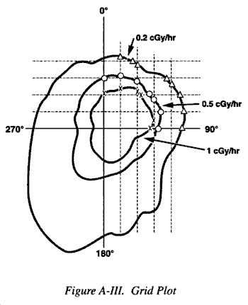
d. The principle objective in an area survey is to establish the location and radiation levels associated with one or more isodose rate lines. An isodose rate line is a plotted contour line that depicts the location of some uniform level of radiation or radioactive contamination.
e. Two methods of plotting isodose rate lines are illustrated in Figures A-II and A-III. The radial plot is the simpler of the two and can be done quickly with minimum personnel. The grid system is more accurate, but it is time consuming and requires a large number of personnel to cover a large area. In practice, both methods might be employed using the best features of each system. The radial plot is used to establish the isodose rate line boundaries and the grid system is used to plot heavily contaminated areas in detail. Care must be used in selecting a focal point for a radial plot as the entire contamination area may be missed. Whatever system is used, the following rules must be observed:
(1) Isodose rate lines must always close.
(2) Isodose rate lines cannot cross each other.
(3) Isodose rate lines can cross a survey line only at a data point.
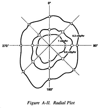
f. To perform an area survey, personnel move into an area until the radiation level established as a guide in the preparatory phase is reached. That point is then designated on a map. Following the readout from the radiation instrument, the isodose rate line for that radiation level is followed to the left or right as terrain dictates until the isodose rate line closes, i.e., a survey team having moved to its right eventually returns to the location in which the first reading was taken and plotted. Having completed this isodose rate line, higher or lower levels of activity are selected and other isodose rate lines are established.
g. The report of an area survey should include the following information: the name of survey team members, the date and time that the survey was performed, the type of radiation detection equipment used in the survey, and other remarks that may be helpful in evaluating the attached survey plot.
h. While area surveys are generally considered as passive surveys, i.e., actions taken based on survey results may be deferred for hours or days, personnel surveys are active, in that actions taken to remove contaminated materials from body surfaces are usually taken immediately. Because they are active surveys, the locations of the contamination on body surfaces are not usually plotted as is the case in area surveys. Instead, contaminated areas are identified with tapes, dyes, magic markers, etc., on the body surface itself.
i. In performing a personnel survey, the individual to be monitored stands, with legs spread and arms extended. The radiation monitor begins the survey at the head, subsequently surveying the upper trunk, arms, lower trunk and legs. The individual being surveyed is asked to do an about face, and the procedure is repeated. As in area surveys, care must be taken not to permit the detector probe to touch any potentially contaminated surfaces. When a contaminated area is identified, it is marked. If it is suspected that contamination may have entered a body opening or wound, swabs may be used to collect surface material. These swabs may then be checked with a radiation detector.
j. Personnel survey records should indicate the name of the individual surveyed, the sites and levels of any activity detected, and the nature of any instructions given to the contaminated individual concerning decontamination procedures.
k. Equipment/material surveys are performed in a manner similar to that used for area surveys. Hand sketches of the object to be surveyed are prepared. Surveying begins at the lower and outer surface of the object to be surveyed and progresses in an upward direction until the object is completely surveyed. Areas of contamination and levels of activity identified are noted on the sketch.
l. Survey records developed from equipment/material monitoring are similar in their information content to those prepared for area surveys.